Case 07
Syncope
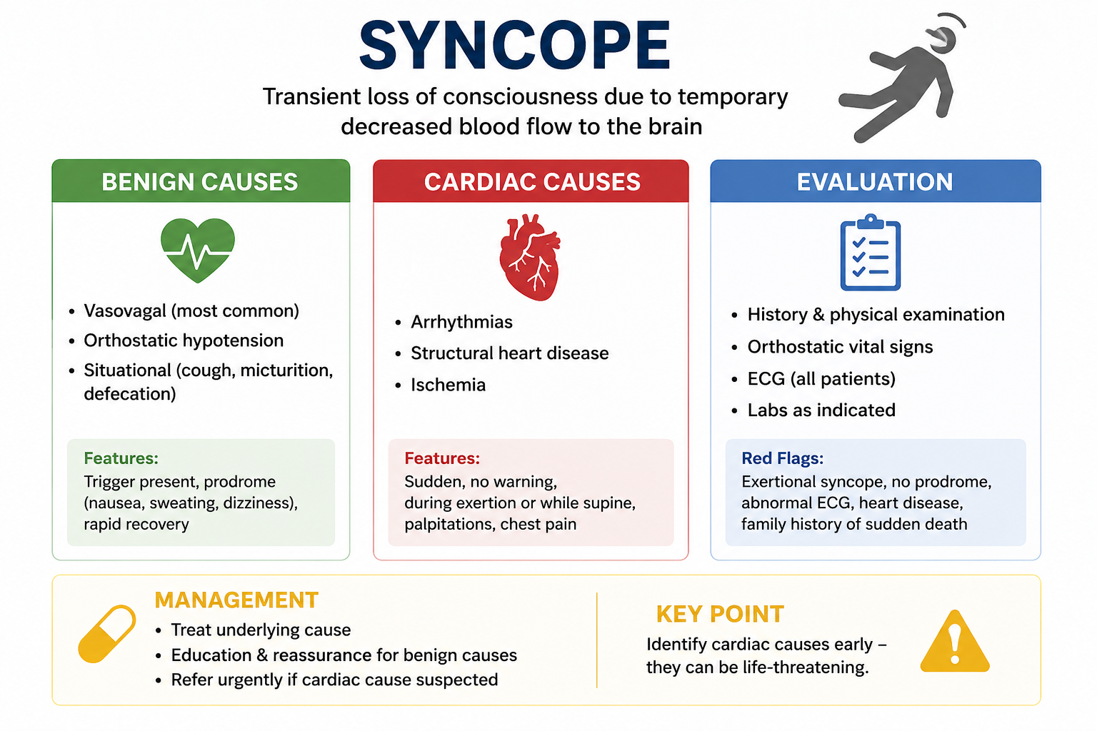
SYNCOPE: CARDIAC VS BENIGN CAUSES
Introduction
Welcome to today’s episode.
Syncope is a common presentation in family medicine and emergency medicine and an important topic in medical licensing examinations, including the MCCQE1, CCFP, Therapeutic Decision-Making Exam, USMLE, PLAB, AMC, and other international examinations.
Most episodes of syncope are benign. However, the clinician’s most important responsibility is recognizing patients whose loss of consciousness may be caused by a dangerous cardiac condition, significant hemorrhage, pulmonary embolism, or another serious disorder.
Definition
Syncope is a sudden, brief, transient loss of consciousness caused by temporary global cerebral hypoperfusion. It is associated with loss of postural tone and is followed by spontaneous and complete recovery.
The three major categories are:
Reflex or vasovagal syncope
Orthostatic hypotension
Cardiac syncope
Common Causes
- Vasovagal Syncope
Vasovagal syncope is the most common cause of syncope.
Typical triggers include emotional stress, pain, fear, prolonged standing, heat exposure, dehydration, seeing blood, and venipuncture.
A prodrome is common. Patients may experience nausea, sweating, a warm sensation, lightheadedness, or blurred or tunnel vision before losing consciousness.
- Orthostatic Syncope
Orthostatic syncope occurs when blood pressure falls excessively after standing.
Common causes include dehydration, blood loss, diuretics, antihypertensive medications, autonomic dysfunction, diabetes, Parkinson disease, and older age.
Orthostatic hypotension is generally defined as a sustained decrease within three minutes of standing of:
Systolic blood pressure of 20 mmHg or more
or
Diastolic blood pressure of 10 mmHg or more
- Cardiac Syncope
Cardiac syncope is particularly important because the underlying cause may be life-threatening.
Potential arrhythmias include ventricular tachycardia, severe bradycardia, high-grade AV block, sick sinus syndrome, long-QT syndrome, and pre-excitation syndromes.
Structural cardiac causes include severe aortic stenosis, hypertrophic cardiomyopathy, severe heart failure, and pulmonary hypertension.
Cardiac syncope may occur suddenly without warning, during exertion, while the patient is supine, or in association with palpitations. It should also be considered carefully in patients with known structural heart disease.
Other Serious Causes of Collapse
Other important conditions should remain in the differential diagnosis.
These include pulmonary embolism, acute myocardial infarction, major internal hemorrhage, ruptured abdominal aortic aneurysm, ectopic pregnancy, hypoglycemia, seizure, stroke in selected presentations, and drug or alcohol intoxication.
Risk Factors for Serious Syncope
Features that increase concern include older age, known heart disease, heart failure, previous myocardial infarction, an abnormal ECG, family history of sudden cardiac death, syncope during exertion, syncope while supine, palpitations immediately before the event, significant anemia or bleeding, and recurrent unexplained episodes.
Clinical Presentation
A practical way to assess syncope is to determine what happened before, during, and after the episode.
Before the Event
Ask whether the patient was standing, exercising, experiencing pain or emotional stress, or having palpitations, chest pain, shortness of breath, nausea, or sweating.
During the Event
If possible, obtain a witness history.
Ask about the duration of unconsciousness, skin colour, abnormal movements, injuries, and tongue biting.
After the Event
Simple syncope usually results in relatively rapid recovery.
Prolonged confusion following the event makes seizure or another neurological condition more likely.
SOAP APPROACH
Subjective Assessment
Ask about the patient’s position immediately before the event, possible triggers, prodromal symptoms, duration of unconsciousness, palpitations, chest pain, shortness of breath, exertional symptoms, previous episodes, cardiac history, neurological symptoms, bleeding, possibility of pregnancy, medications, alcohol or recreational drug use, and family history of sudden death.
A witness history should be obtained whenever possible.
Objective Assessment
Check the patient’s blood pressure, heart rate, respiratory rate, oxygen saturation, and temperature.
Orthostatic Vital Signs
When clinically appropriate, measure the blood pressure and pulse while the patient is lying down and again after standing.
Cardiovascular Examination
Assess the pulse and rhythm and listen carefully for murmurs.
Look specifically for evidence of aortic stenosis, heart failure, and impaired peripheral perfusion.
Neurological Examination
Assess mental status, cranial nerves, strength, sensation, and coordination.
A persistent focal neurological deficit is not typical of uncomplicated syncope and requires further evaluation.
Investigations
12-Lead ECG
An ECG is an important part of the initial assessment of a patient presenting with syncope.
Look for bradycardia, AV block, QT prolongation, ischemic changes, ventricular arrhythmias, pre-excitation, and other conduction abnormalities.
Point-of-Care Glucose
Check glucose particularly when hypoglycemia is possible.
Blood Tests
Laboratory testing should be guided by the history and examination rather than ordered routinely in every patient.
Depending on the clinical presentation, consider:
CBC for anemia or bleeding
Electrolytes
Creatinine
Troponin when cardiac ischemia is suspected
Beta-hCG when pregnancy is possible
D-dimer in selected patients when pulmonary embolism is suspected
Further Cardiac Investigation
Depending on the clinical presentation, further investigations may include Holter monitoring, extended ambulatory monitoring, an event monitor, echocardiography, exercise testing, or an implantable loop recorder.
Assessment: Benign or Potentially Cardiac?
Features Suggesting Vasovagal Syncope
A clear trigger
Prolonged standing
Warm environment
Nausea
Sweating
Lightheadedness
Young, otherwise healthy patient
Normal ECG
Rapid recovery
Features Suggesting Cardiac Syncope
Syncope during exertion
Syncope while supine
Sudden collapse without warning
Palpitations before collapse
Known structural heart disease
Abnormal ECG
Family history of sudden cardiac death
Management
Vasovagal Syncope
Most patients with uncomplicated vasovagal syncope do not require medication.
When the diagnosis is clear, explain the benign mechanism and teach the patient how to recognize and respond to future episodes.
Encourage adequate hydration, avoidance of known triggers and prolonged standing, and slow changes in position.
When the prodrome begins, the patient should sit or lie down immediately.
Physical counter-pressure manoeuvres may also help. Examples include leg crossing, hand gripping, arm tensing, and squatting.
Medication is rarely necessary for ordinary vasovagal syncope.
Orthostatic Hypotension
The first step is identifying and correcting the underlying cause.
Review medications carefully, particularly diuretics, alpha-blockers, vasodilators, and excessive antihypertensive therapy.
Encourage adequate hydration when appropriate.
Acute Volume Depletion
If clinically significant dehydration or hypotension requires emergency treatment, intravenous fluids may be necessary.
0.9% Normal Saline
500 to 1000 mL IV bolus
or
Lactated Ringer’s
500 to 1000 mL IV bolus
Reassess after each bolus. Smaller volumes should be considered in patients at risk of fluid overload.
Persistent Symptomatic Orthostatic Hypotension
Selected patients with persistent symptoms may require specialist-guided pharmacological treatment.
Midodrine
Typical starting dose:
2.5 mg orally three times daily
This may be titrated to:
5 to 10 mg orally three times daily
Avoid dosing close to bedtime because of the risk of supine hypertension.
Fludrocortisone
Typical starting dose:
0.1 mg orally once daily
Monitor blood pressure, potassium, edema, and signs of heart failure.
Medication is not first-line treatment for ordinary vasovagal syncope.
Suspected Cardiac Syncope
Patients with suspected cardiac syncope generally require urgent emergency or specialist assessment.
The goal is not simply to treat the fainting episode. The underlying cardiac cause must be identified.
Symptomatic Bradycardia
If syncope is associated with clinically significant symptomatic bradycardia in an emergency setting:
Atropine
1 mg IV
Repeat every 3 to 5 minutes if required.
Maximum total dose:
3 mg
If atropine is ineffective, urgent pacing and advanced cardiac life-support management may be required.
Ventricular Tachycardia
Syncope caused by ventricular tachycardia is a medical emergency.
If the patient has unstable ventricular tachycardia with a pulse, immediate synchronized cardioversion is generally required.
Cardioversion should not be delayed while attempting routine outpatient medication management.
Hypoglycemia
If the episode is caused by hypoglycemia and the patient is conscious and able to swallow:
Give approximately 15 to 20 grams of oral fast-acting glucose.
Recheck the glucose after approximately 15 minutes.
For severe hypoglycemia when the patient cannot safely take oral glucose, treatment may include intravenous dextrose or:
Glucagon
1 mg IM or SC
Family Medicine Office Management
When a patient presents after an episode of syncope, begin by determining whether the event represents true syncope.
Check vital signs, orthostatic blood pressure when appropriate, blood glucose, and an ECG.
Look specifically for cardiac symptoms, bleeding, pregnancy, medication-related causes, and neurological abnormalities.
The final decision is whether the patient can be safely investigated and managed as an outpatient or requires urgent hospital assessment.
When to Send the Patient to the Emergency Department
Urgent assessment is appropriate when syncope occurs during exercise or while supine.
Other concerning findings include chest pain, significant shortness of breath, persistent hypotension, significant bradycardia, significant tachyarrhythmia, an abnormal ECG concerning for serious cardiac disease, known severe structural heart disease, suspected major bleeding, suspected pulmonary embolism, persistent neurological deficits, or recurrent unexplained syncope with concerning features.
Red Flags
Pay particular attention to:
Exertional syncope
Supine syncope
No prodrome
Palpitations immediately before syncope
Chest pain
Shortness of breath
Abnormal ECG
Structural heart disease
Family history of sudden cardiac death
Persistent hypotension
Differential Diagnosis
Not every transient loss of consciousness is syncope.
Important alternatives include seizure, hypoglycemia, intoxication, vertigo, mechanical falls, and psychogenic pseudosyncope.
Important Points for Medical Examinations
Vasovagal syncope is the most common cause of syncope.
A typical prodrome consisting of nausea, warmth, sweating, and lightheadedness supports a vasovagal cause.
Syncope occurring during exercise should be considered potentially cardiac until adequately evaluated.
An ECG is an important component of the initial syncope assessment.
Routine CT brain imaging is generally unnecessary for uncomplicated syncope in the absence of neurological findings or significant head injury.
Prolonged confusion following the event suggests seizure rather than uncomplicated syncope.
Always review the medication list when evaluating orthostatic hypotension.
Remember that cardiac syncope may be the first warning sign of a potentially fatal arrhythmia.
Conclusion
The most important part of evaluating syncope is determining why the patient temporarily lost consciousness.
Confirm that the episode represents true syncope, assess vital signs, obtain an ECG, and look for features suggesting vasovagal or orthostatic syncope. At the same time, actively search for cardiac red flags and other serious causes.
A patient with a clear trigger, typical warning symptoms, and rapid recovery is more likely to have benign vasovagal syncope.
In contrast, sudden collapse during exertion, while lying down, or without warning should raise immediate concern for a cardiac cause and prompt appropriate urgent evaluation.
This content is for educational purposes only and does not constitute medical advice or clinical guidance. Read the full disclaimer.