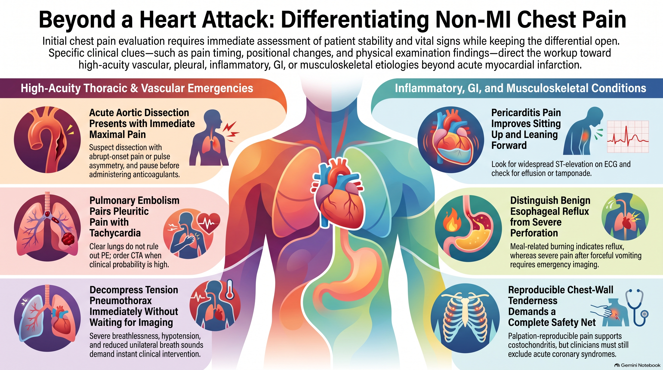
Chest Pain Beyond a Heart Attack: Six Clinical Challenges
A TopMedCast case-based episode
Introduction
A woman develops sharp chest pain after a long journey. A man describes pain that reached its worst intensity in seconds. A young adult feels better sitting forward. Another patient says the discomfort started after dinner.
All have chest pain. Their investigations, treatment and urgency will be very different.
Welcome to TopMedCast. In this episode, we explore six causes of chest pain beyond myocardial infarction: pulmonary embolism, aortic dissection, pericarditis, pneumothorax, oesophageal disease and musculoskeletal pain.
For each case, ask yourself three questions: What is the dangerous diagnosis? Which findings change the probability? And what needs to happen next?
These are fictional educational cases. In practice, assessment and treatment must follow the patient’s condition and local protocols.
Before the Cases: The First Assessment
Before deciding what is causing the pain, decide whether the patient is stable.
Look for respiratory distress, hypoxaemia, hypotension, altered consciousness or poor peripheral perfusion. Obtain vital signs, assess the heart and lungs, and arrange an early ECG. Use troponin testing and repeat assessment when indicated by the presentation and the local chest-pain pathway.
Keep the differential open. Sharp pain does not automatically mean a benign condition. A normal initial ECG does not close the assessment. And an elevated troponin indicates myocardial injury without, by itself, establishing its cause.
Now, our first patient.
Case One: The Journey That Changed the Diagnosis
A 42-year-old woman presents with right-sided chest pain that becomes worse when she breathes deeply. It started that morning, two days after a nine-hour flight.
She feels slightly breathless but believes she has pulled a muscle carrying luggage.
Her heart rate is 112 beats per minute, respiratory rate 24, blood pressure 124 over 78, and oxygen saturation 94% on room air. Her lungs sound clear. On further questioning, she mentions new discomfort in her left calf.
She also takes an oestrogen-containing contraceptive.
What should move to the top of your differential?
Pulmonary embolism.
The concerning combination is pleuritic pain, unexplained tachycardia, breathlessness and possible deep-vein thrombosis, with relevant thrombotic risk factors.
Clear lungs do not explain away those findings.
Choosing the Investigation
Start by estimating clinical probability using a validated approach.
PERC can help avoid testing in appropriately selected patients who already have a low clinical probability. This patient is not PERC-negative.
D-dimer is useful within a low or intermediate probability pathway. A positive result does not diagnose pulmonary embolism. When clinical probability is high, proceed to appropriate imaging rather than using D-dimer to seek reassurance. CT pulmonary angiography is commonly used; a ventilation–perfusion study may be suitable in selected circumstances. acc.org
In this case, CT pulmonary angiography confirms pulmonary emboli.
What Happens Next?
Confirmation starts a second assessment: how dangerous is this embolism for this patient?
Assess haemodynamics, oxygen requirements, right-ventricular function, relevant biomarkers, bleeding risk and suitability for outpatient care.
Anticoagulation is central for eligible patients. Selected low-risk patients can receive outpatient treatment through an established pathway, while patients with instability or cardiopulmonary deterioration need hospital care and urgent assessment for advanced treatment. Thrombolysis is not routine for every pulmonary embolism. ahajournals.org
The clinical lesson: Pleuritic pain with tachycardia deserves an explanation, even when the patient looks relatively comfortable.
Case Two: The Pain That Was Worst at the Beginning
A 67-year-old man with longstanding hypertension develops severe central chest pain while eating breakfast.
When asked how quickly it became severe, he answers, “Immediately. It was the worst right from the start.”
The pain extends between his shoulder blades. He briefly felt that his left leg was weak.
His right-arm blood pressure is 190 over 100. His left radial pulse feels weaker than the right. A soft early diastolic murmur is heard.
Which detail should stop you from treating this as routine coronary chest pain?
Several should: abrupt maximal pain, possible malperfusion, pulse asymmetry and a possible aortic regurgitation murmur.
Together, they raise concern for acute aortic dissection.
The patient does not need to use the word “tearing.” The timing and associated findings matter more than one descriptive adjective.
Investigation and Immediate Treatment
Arrange emergency specialist involvement and urgent aortic imaging. CT angiography is generally the initial investigation when feasible; transoesophageal echocardiography can be useful when the patient cannot safely undergo CT.
Provide analgesia and monitored treatment to reduce aortic wall stress. In hypertensive patients without contraindications, intravenous beta-blockade is the initial approach. Additional vasodilation may follow if needed. Targets must preserve organ perfusion; hypotension requires a different response.
A dissection involving the ascending aorta—Stanford type A—requires emergency surgical assessment. An uncomplicated type B dissection usually begins with medical management, while rupture, malperfusion or other complications may require intervention. ahajournals.org
The Dangerous Diagnostic Shortcut
Suppose this patient also has ECG changes or an elevated troponin.
That does not remove dissection from consideration. A dissection can compromise coronary blood flow.
When dissection is strongly suspected, pause before reflexively administering anticoagulation or thrombolysis. Those treatments may cause serious harm in this setting.
The clinical lesson: Ask, “Was the pain severe immediately, or did it build?” That question can change the entire pathway.
Case Three: The Patient Who Cannot Lie Flat
A 29-year-old man presents with sharp central chest pain several days after a respiratory illness.
The pain worsens with deep inspiration and lying flat. He feels more comfortable sitting upright and leaning forward.
His blood pressure is normal. He has a low-grade temperature and no clinical signs of heart failure.
His ECG shows widespread ST-segment elevation with PR-segment depression.
What diagnosis best brings these findings together?
Acute pericarditis.
The positional and pleuritic pain supports the diagnosis, while the ECG provides additional evidence. However, the tracing must be interpreted in context: ST elevation always requires careful consideration of acute coronary occlusion and other causes.
What Else Should You Check?
Assess inflammatory markers, renal function and troponin, and obtain echocardiography to evaluate ventricular function and look for an effusion.
An elevated troponin may indicate associated myocardial involvement and should change the assessment.
For uncomplicated acute pericarditis, treatment commonly includes aspirin or an NSAID plus colchicine, with attention to contraindications, interactions and gastroprotection. Restrict strenuous exercise until clinical remission and arrange follow-up. Corticosteroids are reserved for selected indications rather than used routinely as first-line treatment. acc.org
When Does This Become an Admission?
Look for high-risk features: significant fever, a subacute course, a large effusion, tamponade, myocardial involvement or an inadequate response to initial treatment. Immunosuppression, trauma and anticoagulation also affect disposition.
Now imagine that the patient becomes hypotensive and increasingly breathless. His jugular venous pressure rises, and echocardiography shows an effusion with features of tamponade.
The priority has changed. He needs urgent expert assessment for drainage and haemodynamic support, not simply stronger anti-inflammatory treatment. jacc.org
The clinical lesson: Recognizing pericarditis is only the beginning. Assess for myocardial involvement, effusion and tamponade.
Case Four: Sudden Pain in a Previously Healthy Patient
A 23-year-old man develops sudden left-sided chest pain and breathlessness while walking.
He has no trauma history. He smokes and has no known lung disease.
His oxygen saturation is 95% on room air. Blood pressure is normal, but breath sounds are reduced on the left.
A chest radiograph demonstrates a left pneumothorax.
Does every pneumothorax require a chest drain?
Management depends on symptoms, physiological stability, underlying lung disease and the available care pathway.
Selected adults with a primary spontaneous pneumothorax who have minimal symptoms and no physiological compromise may be managed conservatively, even when the pneumothorax is radiographically large. Others need ambulatory treatment, aspiration or drainage.
Patients with underlying lung disease require particular caution because their respiratory reserve may be limited. brit-thoracic.org.uk
The Emergency Variation
Now change the case.
The patient becomes severely breathless, hypotensive and confused. Breath sounds are markedly reduced on one side.
Suspect tension pneumothorax.
In an unstable patient with a convincing clinical presentation, do not wait for radiographic confirmation. Arrange immediate decompression by a trained clinician according to the emergency protocol, followed by definitive pleural management.
Tracheal deviation is a late and unreliable sign. Waiting for it can delay treatment.
After the Acute Event
Discuss smoking cessation, recurrence risk and follow-up imaging. Give explicit instructions to return immediately for worsening breathlessness or chest pain.
Air travel must wait until appropriate radiographic resolution and the recommended interval afterward. Scuba diving requires specialist advice because recurrence underwater can be fatal. Thorax
The clinical lesson: Stability guides investigation. Suspected tension physiology demands immediate action.
Case Five: “It Must Be Heartburn”
A 51-year-old woman reports intermittent burning discomfort behind her sternum.
It commonly follows large meals, worsens when she lies down and is sometimes accompanied by a sour taste.
She has started avoiding late dinners because they trigger symptoms.
That pattern suggests gastro-oesophageal reflux. However, her cardiovascular risk and the characteristics of the current episode still require assessment.
A meal-related pattern or improvement with antacid treatment cannot independently establish that chest pain is non-cardiac.
When Reflux Is the Working Diagnosis
Once an appropriate assessment has addressed urgent cardiopulmonary causes, ask about alarm symptoms.
Does food stick? Is swallowing painful? Has she lost weight unintentionally? Is there gastrointestinal bleeding or persistent vomiting?
Chest discomfort can occur with reflux, but those associated features require further evaluation. NIDDK
Without alarm features, management may include avoiding meals close to bedtime, identifying individual triggers, weight management when relevant and an appropriate trial of acid suppression.
Persistent symptoms should prompt reassessment of the diagnosis and consideration of further investigation.
When the Oesophageal Story Is an Emergency
Consider a different history: severe chest pain begins immediately after repeated forceful vomiting.
The patient is tachycardic, distressed and becoming unwell.
That is not a routine reflux presentation. Consider oesophageal perforation and arrange emergency hospital evaluation, urgent imaging and surgical consultation. Keep the patient nil by mouth while resuscitation and specialist-directed treatment proceed.
Other oesophageal disorders, including motility disorders, can also cause chest pain. Investigating them follows appropriate exclusion of urgent causes.
The clinical lesson: “Oesophageal chest pain” covers conditions with very different levels of urgency. The sequence of events matters.
Case Six: The Tender Chest Wall
A 38-year-old man develops anterior chest discomfort after moving heavy furniture.
The pain increases when he twists, reaches overhead or takes a deep breath.
Pressing over several costochondral junctions reproduces the discomfort he came in with. There is no redness, warmth or focal swelling.
He has no breathlessness, syncope, fever or exertional pressure. His vital signs are normal.
After an appropriate assessment, the working diagnosis is costochondritis.
Why the Examination Helps
Matching the presenting pain with palpation and movement supports a chest-wall source. Lack of swelling also fits costochondritis.
Nevertheless, reproducible tenderness does not independently rule out acute coronary syndrome. Its significance depends on the whole presentation, including risk factors and accompanying symptoms.
Management commonly includes explanation, temporary modification of provoking activities, local heat and suitable analgesia. A topical NSAID may be useful; oral treatment requires consideration of gastrointestinal, renal and cardiovascular risks. Persistent symptoms may benefit from stretching or physiotherapy. AFP
Reassurance Needs a Safety Net
Explain why the assessment supports a musculoskeletal diagnosis and what recovery may look like.
Then explain what should trigger reassessment: new breathlessness, fainting, exertional pressure, fever, a changing pain pattern or symptoms that persist despite treatment.
Revisit the diagnosis when the course is unexpected.
The clinical lesson: Chest-wall tenderness is useful evidence. Safe reassurance comes from the complete assessment and a clear follow-up plan.
Bringing the Six Cases Together
Each case had a clue that redirected attention.
For pulmonary embolism, it was pleuritic pain with unexplained tachycardia and thrombotic risk.
For aortic dissection, it was pain that was maximal immediately, accompanied by possible malperfusion.
For pericarditis, it was positional pain with supporting ECG findings—and the need to check for complications.
For pneumothorax, it was sudden breathlessness with unilateral examination findings.
For oesophageal disease, it was the relationship to meals, swallowing or vomiting.
For musculoskeletal pain, it was the match between symptoms, movement and palpation within a reassuring overall assessment.
The challenge is to use those clues without allowing one finding to end the reasoning too early.
Closing
Chest pain assessment begins with urgency: Is the patient stable? What dangerous conditions remain possible? Which investigation or treatment will change the outcome now?
Then comes precision—choosing the right test, recognizing when treatment cannot wait, and knowing when reassurance is justified.
You’ve been listening to TopMedCast: Chest Pain Beyond a Heart Attack—Six Clinical Challenges.
Explore more clinical reasoning episodes at TopMedCast.com, and practise applying your knowledge at TopQBank.com.
This content is for educational purposes only and does not constitute medical advice or clinical guidance. Read the full disclaimer.