Key points
Heart Failure: Six Cases Behind the Breathlessness
Welcome to TopMedCast, where we turn everyday clinical presentations into practical learning for clinicians. For exam-style questions that help you apply your knowledge, visit TopQBank.com.
Today, we explore heart failure through six clinical cases—from the patient who has quietly stopped climbing stairs to the patient struggling to breathe in front of you.
Heart failure can present with pulmonary congestion, peripheral oedema, fatigue or declining exercise tolerance. Some patients have a severely weakened left ventricle. Others have a preserved ejection fraction but elevated filling pressures.
Our challenge is to recognise the syndrome, identify its cause, assess urgency and choose treatment that improves both symptoms and long-term outcomes.
These fictional cases are for clinical education. Individual prescribing requires assessment of blood pressure, renal function, potassium, comorbidities and local guidance.
Case One: “I Thought I Was Just Getting Older”
A 66-year-old man attends with three months of increasing breathlessness.
He previously walked several blocks without difficulty. Now he stops halfway up a flight of stairs. His shoes feel tighter, and he has started sleeping on three pillows.
He has hypertension, type 2 diabetes and a previous myocardial infarction.
His blood pressure is 138 over 82. His pulse is 94 and regular. Oxygen saturation is 95% on room air. Examination reveals elevated jugular venous pressure, bibasal crackles and bilateral pitting ankle oedema.
He describes his symptoms as ageing. The combination suggests heart failure with congestion.
The first decision is whether outpatient investigation is appropriate. Breathlessness at rest, hypoxaemia, chest pain, syncope, hypotension or rapid deterioration would require urgent hospital assessment.
In a stable patient, investigate promptly. An ECG can identify atrial fibrillation, previous infarction or conduction disease. Bloodwork should assess blood count, electrolytes, renal function, liver function, thyroid function and glucose control. Iron studies can identify a clinically relevant contributor to fatigue and exercise limitation.
A chest radiograph may show pulmonary congestion, pleural effusions or an alternative explanation for breathlessness.
BNP or NT-proBNP helps assess the likelihood of heart failure, but interpretation requires context. Levels may rise with atrial fibrillation, renal dysfunction and increasing age. Obesity can produce lower concentrations than expected.
Echocardiography assesses ejection fraction, chamber structure, valve disease and right ventricular function. A normal chest radiograph does not exclude heart failure, and natriuretic peptides should not replace clinical judgment.
His echocardiogram shows a left ventricular ejection fraction of 30%.
He has heart failure with reduced ejection fraction. His previous infarction suggests an ischaemic cause, although further evaluation depends on symptoms and clinical circumstances.
Treatment now has two linked aims: relieve congestion and introduce therapies that improve outcomes. A loop diuretic addresses fluid retention, while disease-modifying treatment is introduced as tolerated.
The lesson from this case is to ask what the patient has stopped doing. Reduced activity can conceal the severity of breathlessness.
Case Two: “But Her Ejection Fraction Is Normal”
A 78-year-old woman presents with exertional breathlessness and intermittent ankle swelling.
She has longstanding hypertension, obesity, diabetes and atrial fibrillation.
Her echocardiogram reports an ejection fraction of 60%. Someone reassures her that her heart is pumping normally.
However, further assessment identifies left atrial enlargement, left ventricular hypertrophy and evidence of elevated filling pressures.
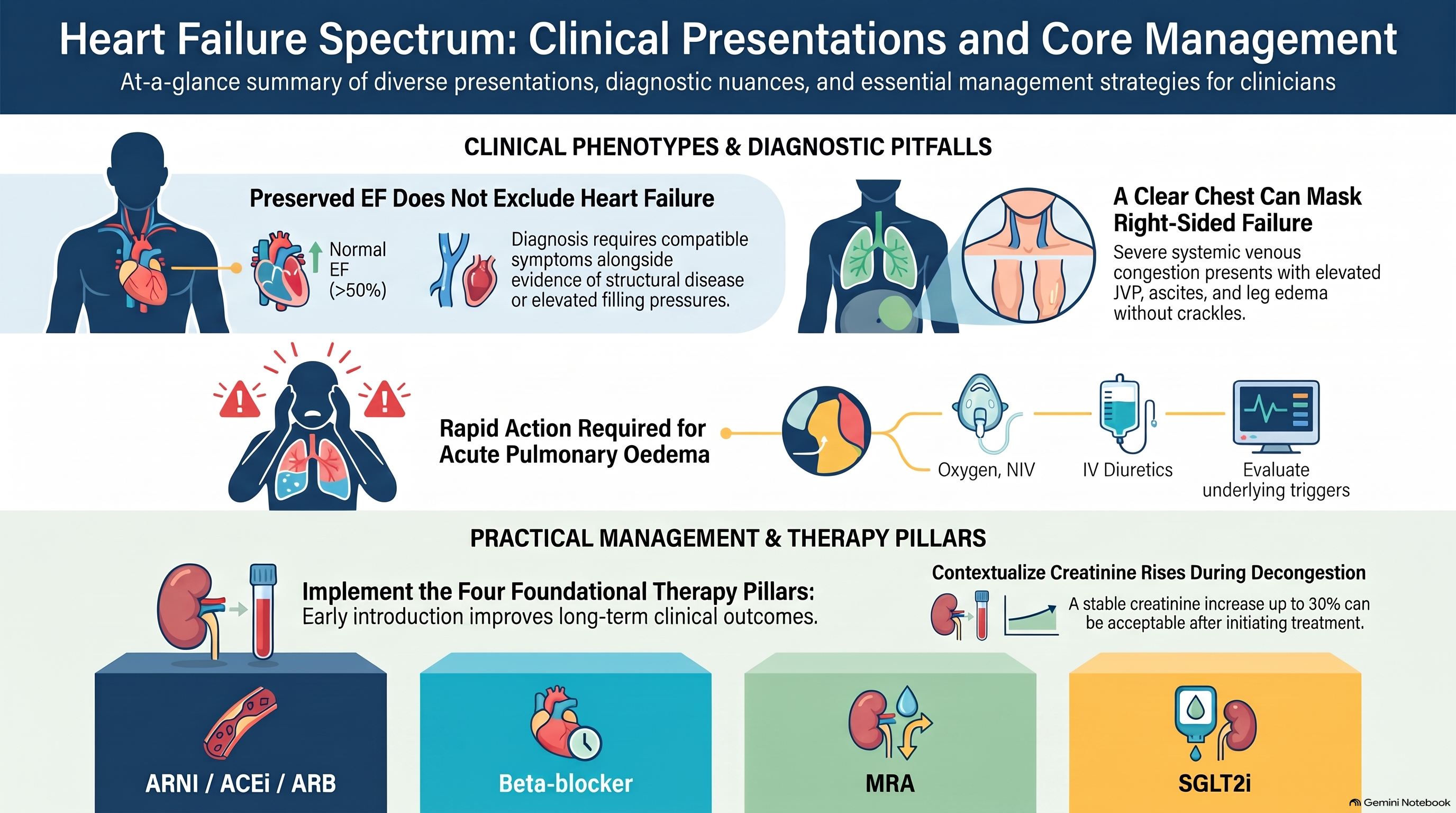
Preserved ejection fraction does not exclude heart failure. Diagnosis requires compatible symptoms and signs together with objective evidence supporting abnormal cardiac structure, function or filling pressures.
Management includes relieving congestion, treating hypertension and atrial fibrillation, and addressing obesity, diabetes, kidney disease and sleep-disordered breathing.
SGLT2 inhibitors reduce heart failure events across the nonreduced ejection fraction spectrum, including in patients without diabetes. Canadian guidance now groups ejection fractions above 40% under heart failure with nonreduced ejection fraction. Additional options include mineralocorticoid receptor antagonists, selected use of sacubitril–valsartan and therapies with GLP-1 receptor agonist activity for appropriate patients with obesity.
The choice depends on the patient’s phenotype, kidney function, potassium, blood pressure and treatment eligibility.
For this woman, follow-up should assess breathlessness, congestion, exercise capacity and medication tolerance.
Her preserved ejection fraction is one part of the assessment. It does not explain away her symptoms.
Case Three: The Patient Who Cannot Finish a Sentence
A 72-year-old man with known heart failure develops rapidly worsening breathlessness overnight.
He arrives sitting upright, sweating and speaking in short phrases.
His respiratory rate is 32. Oxygen saturation is 84% on room air. Blood pressure is 190 over 110. Crackles are audible throughout both lungs.
This is an emergency consistent with acute pulmonary oedema.
If the patient presents in a community clinic, activate emergency transport. Begin immediate assessment and support while arranging transfer.
In a monitored setting, address oxygenation and respiratory distress. Noninvasive ventilation may be needed for persistent hypoxaemia or marked work of breathing. Intravenous loop diuretics treat congestion. Nitrates can be helpful in hypertensive pulmonary oedema when blood pressure and contraindications permit.
Management must follow haemodynamics. Hypotension, poor perfusion or suspected cardiogenic shock requires a different approach.
At the same time, investigate the trigger: acute coronary syndrome, arrhythmia, infection, uncontrolled hypertension, worsening renal function, medication interruption or a new drug promoting fluid retention.
The ECG and targeted investigations help identify these causes. A raised troponin requires interpretation within the overall presentation; it does not automatically establish an acute coronary occlusion.
Routine morphine is not part of standard treatment.
After stabilisation, review how the deterioration developed. Did he run out of medication? Was he taking an NSAID? Did increasing weight or orthopnoea go unreported?
Before discharge, he needs a clear medication plan, follow-up and instructions for recognising worsening congestion.
The lesson is that acute heart failure care includes both immediate stabilisation and finding the reason it happened.
Case Four: Feeling Better Is Only the Beginning
A 59-year-old woman returns after admission with newly diagnosed dilated cardiomyopathy.
Her ejection fraction is 28%. She feels much better after diuresis and asks whether furosemide is the only medicine she needs.
For eligible patients with reduced ejection fraction, treatment includes four foundational medication classes.
The first is an angiotensin receptor–neprilysin inhibitor, usually sacubitril–valsartan, or an ACE inhibitor or ARB when appropriate.
The second is an evidence-based beta blocker, such as bisoprolol, carvedilol or sustained-release metoprolol succinate.
The third is a mineralocorticoid receptor antagonist, such as spironolactone or eplerenone.
The fourth is an SGLT2 inhibitor, such as dapagliflozin or empagliflozin.
Introduce these therapies early as the patient’s condition permits. Avoid unnecessary delays while waiting for one drug to reach its target dose before considering another class.
Prescribing requires several practical safeguards.
Allow at least 36 hours between the last ACE inhibitor dose and the first sacubitril–valsartan dose. Do not combine them.
Start or increase a beta blocker when the patient is clinically stable. Active shock or severe unstable decompensation is not the setting for escalation.
Before starting spironolactone or eplerenone, assess renal function and potassium. A usual eligibility threshold is an estimated GFR above 30 and potassium below 5.0, with ongoing monitoring.
SGLT2 inhibitors provide benefit even without diabetes. Explain volume depletion, genital infections and the uncommon risk of ketoacidosis. Provide instructions about temporary withholding during significant acute illness, prolonged fasting or surgery, following the relevant prescribing guidance.
Arrange early review and bloodwork after relevant medication changes. Adjust treatment according to symptoms, blood pressure, pulse, kidney function and potassium.
After a period of optimised therapy, reassess ventricular function. Persistent dysfunction may warrant evaluation for an implantable defibrillator or cardiac resynchronisation therapy, depending on the full eligibility criteria.
The lesson is that symptom improvement should prompt continued optimisation.
Case Five: Swollen Legs and a Quiet Chest
A 70-year-old man presents with worsening leg swelling, abdominal fullness and early satiety.
He has chronic lung disease and obstructive sleep apnoea.
His lungs have few crackles, but his jugular venous pressure is markedly elevated. He has bilateral pitting oedema, an enlarged liver and ascites.
The absence of prominent crackles does not exclude a cardiac problem.
This pattern raises concern for right-sided heart failure and systemic venous congestion. Echocardiography should assess right ventricular size and function, tricuspid valve disease, estimated pulmonary pressures and associated left-sided disease.
The cause matters. Right ventricular dysfunction may follow left-sided heart disease, pulmonary hypertension, chronic lung disease, thromboembolic disease or valve pathology.
Keep the differential open. Leg oedema can also reflect venous insufficiency, medication effects, renal disease, liver disease or low albumin.
Carefully monitored diuresis can relieve systemic congestion, but hypotension and excessive volume depletion can be particularly problematic in some patients with right ventricular failure.
Pulmonary hypertension requires classification and specialist assessment. Pulmonary arterial hypertension drugs are not routine treatment for every patient with elevated pulmonary pressures.
For this patient, the chronic course allows structured evaluation. Sudden breathlessness, syncope, chest pain or haemodynamic instability would change the urgency.
The lesson is to examine the neck veins and abdomen as carefully as the lungs.
Case Six: The Creatinine Has Increased
A 76-year-old woman with heart failure and chronic kidney disease attends for review after an increase in her diuretic.
Her weight has fallen, her breathing is easier and her oedema has improved.
Her creatinine has increased from 130 to 150 micromoles per litre.
Should treatment automatically be stopped?
First, reassess the patient. Is she still congested? Is she dizzy, hypotensive or volume depleted? What has happened to urine output, potassium and the creatinine trend? Has she developed an intercurrent illness or started an NSAID?
A modest creatinine increase can occur during effective decongestion or after starting renin–angiotensin system treatment.
The clinical meaning depends on the setting. Persistent congestion can itself worsen kidney function. A creatinine increase alone does not establish that a congested patient needs less diuretic.
Conversely, hypotension, dehydration, oliguria, progressive kidney injury or significant hyperkalaemia requires prompt assessment and treatment adjustment.
For ACE inhibitors and ARBs, a stable creatinine increase of up to about 30% can occur after initiation and may be acceptable with monitoring. This is a contextual guide, rather than permission to ignore a deteriorating patient.
In this woman, assess volume status and arrange repeat bloodwork at an interval appropriate to the clinical picture. Decisions should preserve effective heart failure treatment whenever safely possible.
Advanced kidney disease, recurrent hyperkalaemia, persistent congestion or uncertainty about treatment warrants specialist input.
The lesson is to interpret the laboratory result alongside perfusion and congestion.
Helping Patients Recognise Deterioration
Across all six cases, treatment works best when patients understand what to watch for.
Explain the purpose of each medication and agree on a practical monitoring plan.
For patients who can monitor weight, recommend consistent measurements under similar conditions. Provide an individual action plan for rapid weight gain, worsening swelling or increasing breathlessness. Any self-adjustment of diuretics should follow a plan agreed with the treating team.
Review medication access and adherence without blame. Frequent urination, cost, confusion and fear of adverse effects can all interfere with treatment.
Encourage appropriate physical activity and cardiac rehabilitation when clinically stable. Discuss excess dietary sodium, smoking and alcohol. Fluid advice should be individualised; strict fluid restriction is not necessary for every stable patient.
Avoid NSAIDs when possible, and review over-the-counter medicines.
Severe breathlessness, chest pain, fainting, confusion or other signs of acute deterioration require emergency assessment.
Closing Clinical Reflections
These six patients remind us that heart failure has several faces.
A patient may gradually reduce activity before reporting breathlessness. Preserved ejection fraction can coexist with elevated filling pressures. Acute pulmonary oedema requires immediate action. Reduced ejection fraction calls for early foundational treatment. Right-sided congestion may appear mainly in the legs and abdomen. Kidney results require interpretation within the whole clinical picture.
At each encounter, ask: Is the patient congested? Are they adequately perfused? What caused the deterioration? Is their treatment appropriate and tolerated? Do they know when and how to seek help?
Thank you for listening to TopMedCast.
To practise clinical reasoning with exam-style questions, visit TopQBank.com. For more practical clinical discussions, visit TopMedCast.com.
TopMedCast uses AI-assisted content and voice production. This episode supports clinical learning and should be applied alongside current guidance and individual clinical judgment.
This content is for educational purposes only and does not constitute medical advice or clinical guidance. Read the full disclaimer.