The Swollen Leg
Clinical Clues, Diagnostic Pitfalls and Management Decisions
A swollen leg is a presentation, not a diagnosis. One patient needs urgent anticoagulation, another needs antibiotics, and another needs long-term compression and skin care. The challenge is recognising the pattern while avoiding premature conclusions.
In this TopMedCast episode, we follow six patients with leg swelling. For each case, we explore the history, examination, investigations, management and decisions that change the outcome.
The cases are fictional teaching scenarios.
Start With the Pattern
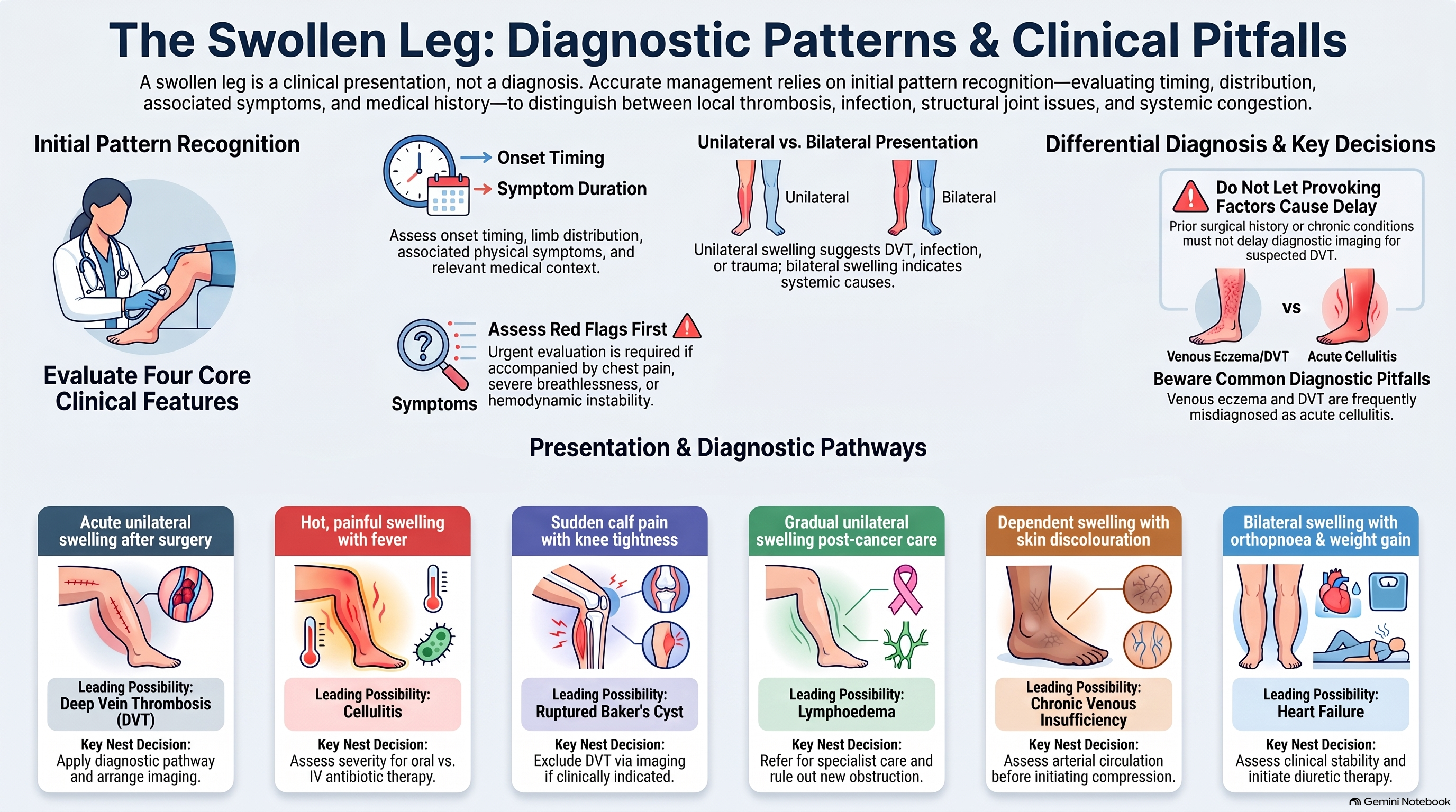
Before focusing on a particular diagnosis, establish four things:
- Timing: Did the swelling develop over hours, days or months?
- Distribution: Is one leg affected, or both? Is the swelling confined to the calf or does it involve the entire limb?
- Associated features: Is there pain, warmth, redness, fever, breathlessness or a change in skin colour?
- Context: Has there been surgery, immobility, trauma, cancer treatment, a medication change or previous thrombosis?
Acute unilateral swelling raises concern for DVT, infection, injury or a ruptured Baker’s cyst. Chronic swelling suggests venous or lymphatic disease. Bilateral swelling broadens the assessment to systemic causes, including heart failure, kidney or liver disease, and medication effects.
These patterns guide the assessment, but they do not establish the diagnosis. Unexplained unilateral swelling deserves prompt evaluation, particularly when accompanied by pain. Swelling with chest pain or breathlessness may indicate a pulmonary embolism and requires urgent assessment. www.nhs.uk
Case 1: The Swollen Calf After Surgery
Deep Vein Thrombosis
The presentation
A 64-year-old woman attends the clinic ten days after a right knee replacement. Her recovery initially went well, but during the last two days her right calf has become increasingly swollen and uncomfortable.
She assumed the swelling was part of the normal postoperative recovery. Today, however, her trousers feel tighter around the calf and walking is more painful.
She has no fever, chest pain or breathlessness. She has been taking her prescribed postoperative thromboprophylaxis.
The assessment
The clinician asks about the onset and progression of swelling, mobility since surgery, missed medication doses, previous DVT or pulmonary embolism, active cancer and bleeding history.
Examination compares both legs. The right calf is larger, with pitting oedema and tenderness. Vital signs are stable.
The key question is whether this is expected postoperative swelling or a new thrombus. Recent surgery increases thrombotic risk, and taking prophylaxis does not completely exclude DVT.
The investigation
Use a validated diagnostic pathway based on clinical probability. In a patient with likely DVT, compression ultrasonography is the priority; a D-dimer alone should not be used to rule out the diagnosis.
D-dimer is often elevated after surgery, so a positive result in this setting is particularly nonspecific. If imaging is delayed, the pathway may recommend interim therapeutic anticoagulation after assessing bleeding risk and contraindications.
A negative initial proximal ultrasound does not always finish the assessment. Depending on the D-dimer result and the pathway used, repeat imaging may be required. NICE recommends repeat proximal ultrasound after six to eight days when the initial scan is negative but the D-dimer is positive. www.nice.org.uk
The result and management
Ultrasound confirms a proximal DVT.
Treatment requires therapeutic anticoagulation, selected according to renal function, bleeding risk, drug interactions, pregnancy status, cancer history and other relevant conditions. Postoperative prophylactic dosing is insufficient treatment for an established proximal DVT.
For many adults with confirmed proximal DVT, anticoagulation continues for at least three months, followed by reassessment of recurrence and bleeding risks. www.nice.org.uk
The diagnostic pitfall
Calling the swelling “normal after surgery” without reassessing a changing clinical picture.
The teaching message
A clear provoking factor should increase suspicion. It should not become an explanation that delays investigation.
Case 2: The Hot, Tender Leg With Fever
Cellulitis
The presentation
A 48-year-old man presents with a red, swollen left lower leg that has worsened over two days. The skin feels hot and painful, and he had chills overnight.
He has type 2 diabetes and frequently develops itchy, cracked skin between his toes. He does not recall an injury.
The assessment
The clinician establishes how quickly the redness is spreading and asks about fever, vomiting, severe pain, recent wounds, water exposure, bites, previous cellulitis and immune suppression.
Examination shows a warm, tender area of erythema extending from the ankle to the lower shin. There is a fissure between the toes.
The skin break provides a possible entry point for infection. Cellulitis commonly causes painful, hot, swollen skin and can spread beyond the original area if untreated. www.nhs.uk
The decision about severity
The clinician must decide whether the patient can be managed with oral treatment and reliable follow-up or needs hospital assessment.
Assess vital signs, mental status, hydration and the extent of infection. Rapid progression, severe systemic illness, haemodynamic instability or concern for deeper infection changes the urgency.
Pain that seems much greater than the visible findings should prompt reassessment rather than reassurance.
The management
Start an antibiotic appropriate for non-purulent cellulitis according to local guidance, allergies and clinical severity. Purulence or an abscess changes management because drainage may be required.
Marking the edge of erythema can help assess progression. Elevation may reduce discomfort and swelling. Treat the toe-web skin disease and discuss foot care.
Severe cellulitis may require intravenous antibiotics and hospital care. www.nth.nhs.uk
Follow-up
Explain what improvement should look like and provide clear instructions for worsening pain, spreading redness or systemic symptoms. Persistent symptoms require reassessment of the diagnosis, treatment and possible complications.
The diagnostic pitfall
Assuming every red leg is cellulitis. Venous eczema, lymphoedema and DVT can produce overlapping findings.
The teaching message
The diagnosis depends on the whole pattern: acute progression, warmth, tenderness, systemic features and a plausible entry point.
Case 3: Sudden Calf Pain in a Patient With Knee Arthritis
Ruptured Baker’s Cyst
The presentation
A 59-year-old woman with knee osteoarthritis develops sudden left calf pain while walking. The calf becomes swollen over the following day.
For several weeks, she had noticed tightness behind the knee, especially when bending it. She has no fever or chest symptoms.
The assessment
A Baker’s cyst is a fluid-filled swelling behind the knee, often associated with an underlying knee disorder. If it ruptures, fluid can track into the calf and cause pain, swelling and redness.
The presentation can closely resemble DVT. A history of knee fullness makes a ruptured cyst plausible, but it does not safely exclude a clot. www.nhs.uk
The investigation
Assess DVT probability and investigate through the appropriate pathway. Ultrasound can assess the veins and may demonstrate a cyst or fluid tracking into the calf.
In this case, imaging excludes DVT and supports a ruptured Baker’s cyst.
The management
Management is usually conservative: relative rest during the painful phase, elevation and suitable analgesia. Review the underlying knee problem and consider further musculoskeletal assessment if symptoms persist or the diagnosis remains uncertain.
The patient should understand that swelling may take time to settle and that worsening symptoms need reassessment. www.nhs.uk
The diagnostic pitfall
Treating a suggestive history as definitive proof. “It sounds like a cyst” is not enough when DVT remains clinically possible.
The teaching message
Some DVT mimics are convincing precisely because they produce the same anatomical symptoms.
Case 4: A Heavy Leg After Pelvic Cancer Treatment
Lymphoedema
The presentation
A 56-year-old woman reports gradually progressive swelling of her left leg following pelvic surgery and lymph-node removal.
The swelling is usually painless, but the leg feels heavy. Her shoe no longer fits comfortably, and the skin has become firmer. She has also had two previous episodes of cellulitis.
The assessment
The history suggests impaired lymphatic drainage. Ask about the timing relative to cancer treatment, progression, functional impact, previous infections and any new pelvic or systemic symptoms.
Examination assesses the distribution of swelling, skin texture, foot involvement and evidence of infection. Early lymphoedema may be soft and pitting; longstanding swelling can become thickened and fibrotic.
Importantly, new or changing swelling in someone with a cancer history still requires consideration of other causes, including thrombosis or obstruction related to malignancy. www.nhs.uk
The management
Refer for specialist lymphoedema assessment. Treatment commonly combines:
- Appropriately fitted compression garments or bandaging.
- Exercise to support muscle-assisted fluid movement.
- Daily skin care.
- Selected manual lymphatic drainage techniques.
- Education about maintaining improvements over time.
This approach controls swelling rather than providing a permanent cure. www.nhs.uk
Preventing complications
Skin care is central. Cracks and wounds increase infection risk, and cellulitis can further damage an already compromised limb.
Teach the patient to seek prompt assessment for new redness, warmth, pain or fever. www.nhs.uk
The diagnostic pitfall
Assuming every episode of increased swelling is simply the patient’s established lymphoedema.
The teaching message
A chronic diagnosis does not explain every new change. Look for superimposed infection, thrombosis or obstruction.
Case 5: Swollen Ankles After a Day on the Feet
Chronic Venous Insufficiency
The presentation
A 67-year-old shopkeeper describes ankle swelling that worsens during long shifts and improves overnight. His legs ache toward the end of the day.
He has visible varicose veins, brown discolouration around the ankles and itchy, scaly skin. Both legs are affected, although the right is worse.
He has previously received antibiotics twice for presumed cellulitis, with little benefit.
The assessment
The dependent pattern, chronicity and skin changes suggest venous hypertension with venous eczema.
Ask about previous DVT, ulcers, bleeding varicose veins, occupational standing and symptoms suggesting arterial disease. Examine the skin, pulses and any wounds.
Chronic venous disease can cause swelling, pigmentation, eczema and ulceration. The skin inflammation may be mistaken for infection, especially during a flare. www.nhs.uk
The management
Encourage regular walking and calf-muscle movement, breaks from prolonged standing and leg elevation when practical.
Compression can improve venous return, but first assess arterial circulation and whether compression is appropriate. Suspected arterial disease requires further assessment before choosing the compression regimen.
Treat dry skin with emollients and active eczema with an appropriate topical corticosteroid course. Assess ulcers and consider vascular referral when indicated. www.nhs.uk
The diagnostic pitfall
Repeatedly prescribing antibiotics for chronic bilateral redness without reconsidering venous eczema.
The teaching message
Duration, dependency and skin changes often distinguish venous disease from an acute infection.
Case 6: Swollen Legs, Weight Gain and Night-Time Breathlessness
Heart Failure
The presentation
A 76-year-old man presents with increasing swelling of both legs over two weeks. His shoes are tight and his weight has increased.
On further questioning, he is breathless walking across the house and now sleeps on three pillows. He recently stopped taking his diuretic because frequent urination was inconvenient.
The assessment
The leg swelling is one part of a wider congestion syndrome.
Assess oxygen saturation, respiratory rate, blood pressure, pulse, weight and signs of impaired perfusion. Examination looks for raised jugular venous pressure, lung crackles and peripheral oedema.
Ask about chest pain, palpitations, infection, medication adherence and medicines that could contribute to fluid retention.
The investigation
Evaluation generally includes an ECG, renal function and electrolytes, and other tests directed by the presentation. Natriuretic peptide testing supports assessment of suspected heart failure, while echocardiography evaluates cardiac structure and function.
Breathlessness at rest, hypoxia or haemodynamic instability requires urgent hospital assessment. A patient with acute deterioration should not wait for routine outpatient investigations. www.nice.org.uk
The management
Treat congestion with an appropriate diuretic regimen and monitor clinical response, renal function and electrolytes. Identify the precipitating factor and review longer-term heart-failure therapy according to the cardiac phenotype and comorbidities.
In this case, discuss why the patient stopped treatment. A practical dosing schedule and clear explanation may improve adherence more effectively than simply repeating the prescription. www.nice.org.uk
The diagnostic pitfall
Treating swollen ankles in isolation while overlooking progressive breathlessness and orthopnoea.
The teaching message
When both legs swell, ask what the heart, kidneys, liver and medications may be contributing.
Bringing the Six Cases Together
Presentation
Leading possibility
Important next decision
Acute unilateral swelling after surgery
DVT
Apply a validated diagnostic pathway and arrange timely imaging
Hot, painful swelling with fever
Cellulitis
Assess severity and suitability for outpatient treatment
Sudden calf pain with preceding knee fullness
Ruptured Baker’s cyst
Exclude DVT when indicated
Gradual swelling after lymph-node treatment
Lymphoedema
Assess for additional causes and arrange specialist care
Dependent swelling with pigmentation and eczema
Venous insufficiency
Assess arterial circulation before compression
Bilateral swelling with orthopnoea and weight gain
Heart failure
Assess stability and treat congestion
Closing Message
The swollen leg becomes easier to assess when we ask a consistent set of questions: How quickly did it happen? Is it one leg or both? What symptoms accompany it? What has changed in the patient’s health, treatment or mobility?
DVT deserves timely investigation, but the differential remains broad. Infection, joint disease, lymphatic damage, venous hypertension and cardiac congestion each leave clues—and recognising those clues leads to the right treatment.
Thank you for listening to TopMedCast. Explore more clinical episodes at TopMedCast.com, then put your knowledge into practice with exam-style questions at TopQBank.com. Keep listening, keep learning, and build confidence one case at a time.
This content is for educational purposes only and does not constitute medical advice or clinical guidance. Read the full disclaimer.