Show notes & transcript
Red Eye: Differential Diagnosis and Quick Management in Family Practice
Welcome to TopMedCast. Today, we’re reviewing the differential diagnosis of red eye and the practical first steps a family physician can take.
Red eye can represent anything from mild allergy to a sight-threatening emergency. The most useful starting questions are: Has vision changed? Is there significant pain? Is there photophobia? Does the patient wear contact lenses?
Ask about trauma, chemical exposure, recent eye surgery or injections, discharge and previous episodes. Document visual acuity in each eye separately. Examine the pupils, cornea, eyelids and eye movements, and use fluorescein when indicated.
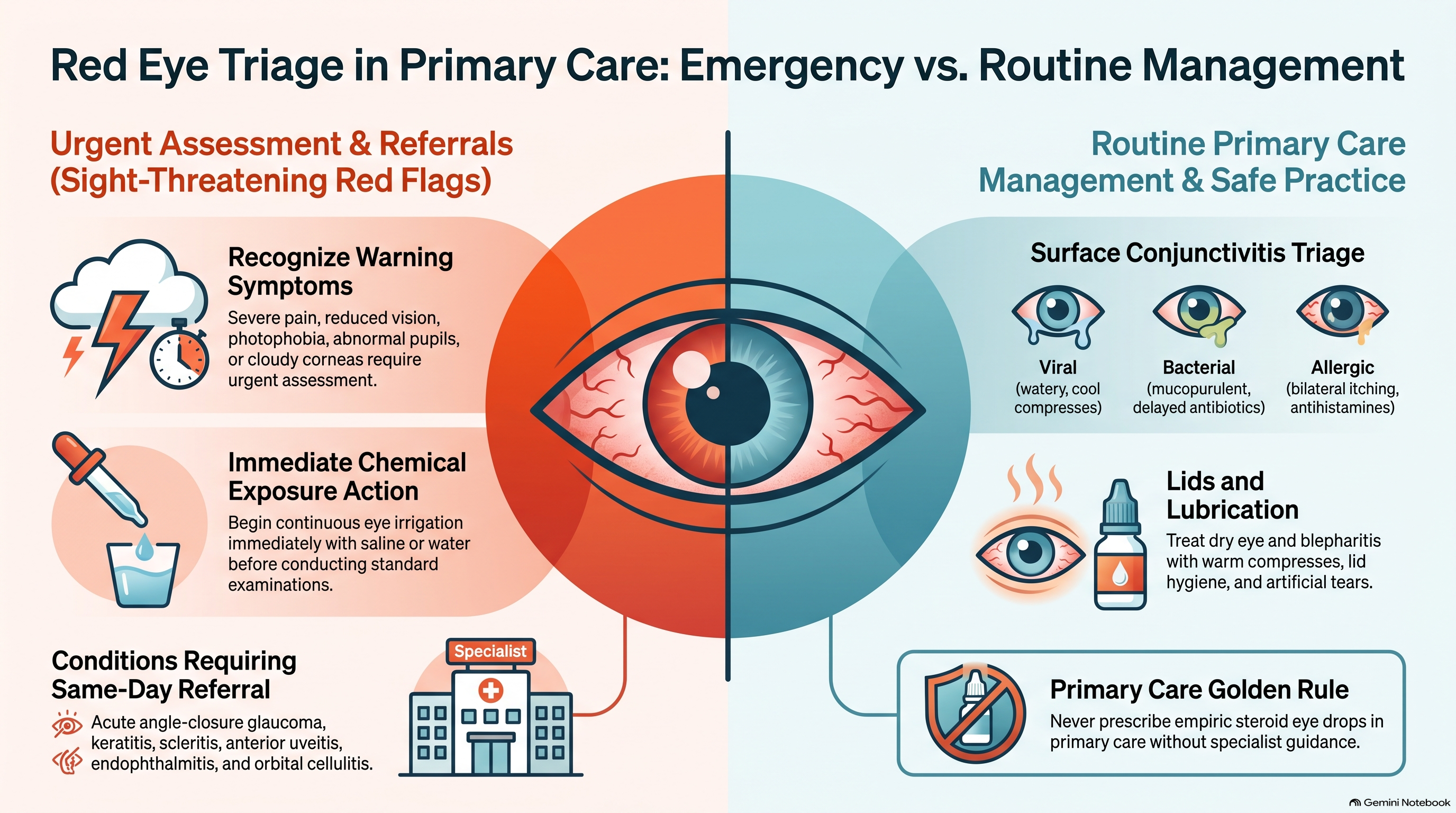
Significant pain, reduced vision, an abnormal pupil or a cloudy cornea requires urgent assessment. Chemical exposure is the exception to the usual examination sequence: start irrigation immediately.
1. Viral conjunctivitis
Think viral conjunctivitis when the eye is watery and gritty, particularly with a recent respiratory infection or contact with someone who has similar symptoms. It may start in one eye and spread to the other.
As a family physician, recommend cool compresses, artificial tears and careful hand hygiene. Advise against sharing towels or eye cosmetics, and stop contact lens use until recovery.
Antibiotics do not treat viral conjunctivitis. Reassess if symptoms worsen, vision changes or significant pain develops.
2. Bacterial conjunctivitis
Mucopurulent discharge and eyelids stuck together after sleep support bacterial conjunctivitis, although symptoms overlap with viral disease.
For mild, uncomplicated cases, observation or delayed prescribing is reasonable. If treatment is appropriate, use a topical antibiotic according to the local formulary. Expect improvement within about 48 hours; reassess persistent or worsening symptoms.
A painful red eye in a contact lens wearer needs careful corneal assessment before being labelled conjunctivitis.
3. Allergic conjunctivitis
Bilateral itching is the strongest clinical clue. Watery discharge, sneezing and seasonal recurrence make allergy more likely.
Start with allergen avoidance, cool compresses and artificial tears. Add a topical antihistamine or a combined antihistamine and mast-cell stabilizer, such as olopatadine or ketotifen, when appropriate.
Avoid routine steroid eye drops in primary care. Marked photophobia or reduced vision requires assessment for another diagnosis.
4. Dry eye
Dry eye commonly causes burning, grittiness and fluctuating blur that may improve with blinking.
Start artificial tears; use preservative-free preparations when drops are needed frequently. Consider lubricating gel or ointment at bedtime. Encourage regular blinking, screen breaks and reduced exposure to smoke or direct airflow.
Review contributing medications and treat associated eyelid disease. Persistent symptoms despite treatment warrant optometry or ophthalmology assessment.
5. Blepharitis
Look for crusting around the lashes, irritated lid margins and blocked meibomian glands.
Recommend warm compresses, gentle lid massage and regular lid cleansing. Add artificial tears for associated dryness. Explain that ongoing care is often needed because symptoms recur.
Persistent anterior blepharitis may require a prescribed antibiotic ointment, while resistant disease or corneal involvement warrants specialist assessment.
6. Subconjunctival haemorrhage
A sharply defined red patch with normal vision and no significant pain usually represents a subconjunctival haemorrhage.
Reassure the patient that an uncomplicated episode generally resolves over one to two weeks. Artificial tears can relieve mild irritation.
Check blood pressure and ask about trauma, anticoagulants and other bleeding. Recurrent episodes may justify further investigation. Do not stop a prescribed anticoagulant solely because of an isolated uncomplicated haemorrhage without assessing the wider clinical situation.
7. Corneal abrasion or superficial foreign body
Sudden pain, tearing and a foreign-body sensation after minor trauma suggest an abrasion. Fluorescein identifies the epithelial defect.
Remove a superficial foreign body only when you have suitable equipment and experience. Provide oral analgesia and lubrication. Topical antibiotic prophylaxis is commonly used, particularly for contaminated injuries; contact lens–associated abrasions require antipseudomonal coverage.
Avoid patching and stop contact lenses until healing. Arrange review within 24 hours for contact lens–related, large or central abrasions, or when symptoms are not improving.
High-velocity injury, suspected penetration or an embedded foreign body requires urgent specialist assessment.
8. Infectious keratitis or corneal ulcer
A painful red eye with photophobia, reduced vision and a white corneal infiltrate should raise immediate concern for keratitis—especially in a contact lens wearer.
Stop contact lenses and arrange urgent same-day ophthalmology assessment. Contact the specialist promptly to coordinate corneal sampling and intensive antimicrobial treatment.
Do not use a routine conjunctivitis regimen as a substitute for keratitis treatment. Do not patch the eye or start topical steroids without ophthalmology advice.
9. Herpes simplex keratitis
A branching dendritic corneal lesion with terminal bulbs suggests herpes simplex epithelial keratitis.
Arrange urgent ophthalmology assessment and coordinate antiviral treatment. Avoid empiric steroid drops, which can worsen epithelial herpetic infection. Steroids have a role in selected deeper herpetic disease only under specialist supervision.
10. Anterior uveitis
Consider anterior uveitis when there is deep aching pain, photophobia, little discharge and redness concentrated around the cornea. A small or irregular pupil and anterior chamber cells support the diagnosis.
Arrange urgent ophthalmology assessment, generally the same day or within 24 hours depending on severity and local access. Provide oral analgesia while arranging care.
Cycloplegic and steroid drops are commonly used after confirmation of the diagnosis and exclusion of corneal infection. Ask about recurrent episodes, inflammatory back pain, psoriasis and inflammatory bowel disease.
11. Episcleritis
Localized superficial redness with mild discomfort and preserved vision suggests episcleritis.
Most uncomplicated cases can be managed with reassurance, cool compresses and artificial tears. An oral anti-inflammatory may help selected patients if there are no contraindications.
Refer if the diagnosis is uncertain, symptoms persist or episodes recur. Significant pain or visual change should prompt reconsideration of scleritis or another serious cause.
12. Scleritis
Severe, deep, boring pain—sometimes worse at night or with eye movement—raises concern for scleritis. The redness may appear deep and violaceous.
Arrange urgent same-day ophthalmology assessment. Provide oral analgesia and assess for associated autoimmune or infectious disease.
Treatment may require systemic anti-inflammatory or immunosuppressive medication. Avoid starting systemic steroids empirically before discussing the diagnosis with the specialist.
13. Acute angle-closure glaucoma
Severe eye pain with blurred vision, halos, headache, nausea or vomiting is an emergency pattern. A hazy cornea and a mid-dilated, poorly reactive pupil strengthen the concern.
Arrange immediate emergency and ophthalmology care. In a setting equipped for acute treatment, coordinate pressure-lowering medication with ophthalmology while checking contraindications. Provide analgesia and an antiemetic.
Definitive treatment commonly involves laser peripheral iridotomy. Do not delay transfer while trying to complete an office-based workup.
14. Endophthalmitis
A red eye with pain and reduced vision following intraocular surgery or an injection should raise concern for endophthalmitis.
Contact ophthalmology immediately and arrange emergency assessment. Specialist treatment usually requires intravitreal antibiotics and sometimes surgery. Topical antibiotics alone are inadequate.
15. Preseptal and orbital cellulitis
Preseptal cellulitis causes eyelid redness and swelling, with preserved vision, normal eye movements and no proptosis.
A well patient with clearly uncomplicated preseptal cellulitis may be treated with oral antibiotics following local guidance, with close review within 24 to 48 hours. Young infants, systemically unwell patients or those whose eye cannot be adequately examined need hospital assessment.
Painful or restricted eye movements, diplopia, proptosis or reduced vision suggest orbital cellulitis. Arrange emergency hospital assessment for intravenous antibiotics, imaging and specialist care.
16. Chemical eye injury
Begin copious irrigation immediately with saline or water if saline is unavailable. Remove contact lenses when feasible and check ocular surface pH.
Continue irrigation until pH normalizes and remains stable after a pause. Assess for retained particles and arrange emergency eye assessment.
Do not attempt to neutralize an acid with an alkali, or an alkali with an acid. Irrigation is the priority.
17. Suspected penetrating injury
High-velocity trauma, an irregular pupil or other signs of globe injury require emergency care.
Apply a rigid eye shield. Avoid pressure, tonometry and manipulation of the eye. Do not remove an embedded object.
Arrange immediate emergency and ophthalmology assessment, with analgesia and antiemetic treatment as appropriate.
18. Hyperacute discharge or neonatal conjunctivitis
Copious, rapidly recurring purulent discharge raises concern for gonococcal infection. Arrange urgent assessment, appropriate specimens and systemic treatment coordinated with specialist and local sexually transmitted infection guidance.
A newborn with a red eye and discharge also needs prompt assessment. Gonococcal, chlamydial and herpetic infections require specific treatment; routine topical drops alone may be insufficient.
Closing
For the family physician, the first decision is whether the patient can safely receive office treatment or needs urgent specialist care.
Treat straightforward surface disease, arrange appropriate follow-up and give clear return precautions. New visual loss, increasing pain, photophobia or worsening swelling should trigger urgent reassessment.
Remember: document vision, examine the cornea, take contact lens use seriously and avoid empiric steroid eye drops in an undifferentiated red eye.
Thank you for listening to TopMedCast.
This content is for educational purposes only and does not constitute medical advice or clinical guidance. Read the full disclaimer.